Ostracod
From Wikipedia, the free encyclopedia
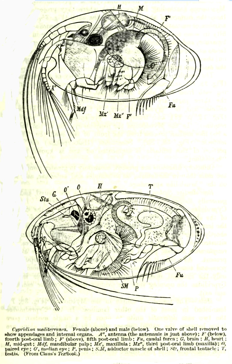
[Photo] Anatomy of Seed Shrimp, Cypridina mediterranea. Source:" A Treatise of zoology". Date 1909. Author E Ray Lankester
Ostracoda is a class of the Crustacea, sometimes known as the seed shrimp because of their appearance. Some 50,000 extinct and extant species have been identified, grouped into several orders.
Ostracods are small crustaceans, typically around one mm in size, but varying between 0.2 to 30 mm, laterally compressed and protected by a bivalve-like, chitinous or calcareous valve or "shell". The hinge of the two valves is in the upper, dorsal region of the body.
Ecologically ostracods can be part of the zooplankton, or (most commonly) they are part of the benthos, living on or inside the upper layer of the sea floor. Many ostracods are also found in fresh water and some are known from humid continental forest soils.
Fossils
Ostracods have a long and well-documented fossil record from the Cambrian to the present day. An outline microfaunal zonal scheme based on both foraminifera and ostracoda was compiled by M. B. Hart (1972).
Ostracods have been particularly useful for the biozonation of marine strata on a local or regional scale, and they are invaluable indicators of paleo-environments because of their widespread occurrence, small size, easily-preservable generally-moulted calcified bivalve carapaces, the valves are a commonly found microfossil.
Morphology
The body of an ostracod is encased by two valves, which together form the duplicature. A distinction is made between the valve (hard parts) and the body with its appendages (soft parts).
Soft parts and ontogeny
The body consists of a cephalon (head), separated from the thorax by a slight constriction. The segmentation is unclear. The abdomen is regressed or absent whereas the adult gonads are relatively large. There are 5-8 pairs of appendages. The branchial plates are responsible for oxygenation.
During the ontogeny the epidermis (containing mesodermal tissue) invaginates ventrolaterally near the cephalon/thorax area. This invagination proceeds upwards and tailwards, until the whole animal is enveloped by a double tissue layer on both sides: this forms the duplicature. The dorsal region never becomes invaginated, and is called the isthmus. The mesodermal tissue in the duplicature develops into the vestibulum. The vestibulum makes contact with the body near the isthmus. It plays a role in oxygenation. In paleo-ecology, the size of the vestibulum can be cautiously interpreted as an environmental indicator. The two double tissue layers surrounding the animal each have an inner and an outer lamella, which surrounds the vestibulum. These lamellae are surrounded by a chitinous cuticle, that is secreted by the epidermal cells.
Like all arthropods, ostracods develop discontinually. Before reaching maturity 8 larval stages (instars) are passed.
Hard parts
The epidermal cells may also secrete calcium carbonate after the chitinous layer is formed, resulting in a chalk layer enveloped by chitin. This calcification is not equally pronounced in all orders. During every instar transition, the old carapace (chitinous and calcified) is rejected and a new, larger is formed and calcified. The outer lamella calcifies completely, while the inner lamella calcifies partially, with the rest remaining chitinous. The partial inner lamella calcification occurs when the ostracod becomes adult. The partial inner lamella calcification is most strongly developed frontally (see electron micrograph). The marginal zone is the area where inner and outer lamella meet, and includes part of the vestibulum. The edge of the marginal zone is called the fused zone, and in this area inner and outer lamella join. The fused zone can contain marginal pore canals. These, along with non-marginal pore canals (that are dispersed evenly along the ostracod's valve) connect the vestibulum to the outer world. The line of concrescence is the visible line between the vestibulum and the fused zone. In many cases, this line is wavering and follows the marginal pore canals. On the inner lamella, a selvage may be present.
http://en.wikipedia.org/wiki/Ostracod
| The text in this page is based on the copyrighted Wikipedia article shown in above URL. It is used under the GNU Free Documentation License. You may redistribute it, verbatim or modified, providing that you comply with the terms of the GFDL. |
|