|
| Query: ciliate | Result: 9th of 22 | |
Protozoa - new scans, #8 - Some more Euplotes - attention, bandwidth intensive
| Subject: | Protozoa - new scans, #8 - Some more Euplotes - attention, bandwidth intensive
| | Poster: | Ralf Schmode (schmode@vossnet.de)
| |

| File size : 97853 bytes
File date : 1998:08:23 09:00:00
Resolution: 793x586
Jpeg process : Baseline
Posted Newsgroups: alt.binaries.pictures.animals
Posted Date: 22 Aug 1998 15:56:03 -0500 |
Protozoa - new scans, #8 - Some more Euplotes - attention, bandwidth intensive
Hello again,
I already showed you a picture of an Euplotes. Euplotes, as I told you,
is a ciliate which has a specialized form of locomotion organs being
called cirri; they have emerged from a process of grouping and
reinforcing the cilia known of ciliates existing earlier in evolution.
The first shot, made in phase contrast illumination, gives you a good
impression of the cirri; compared to the cilia of Paramecium caudatum or
Climacostomum virens the cirri of Euplotes provide much more stiffness.
Indeed, if you watch Euplotes move on the object slide, the cirri are
used to run on the ground rather than swim in the free water. As you can
see, most cirri are located on the outline of the organism. There is an
additional row of cirri pointing towards the middle of the body which is
used for flushing food items into Euplotes' mouth. The mouth itself are
invisible in this shot; however, a lot of greenish food vacuoles being
occupied in algae digestion can be seen.
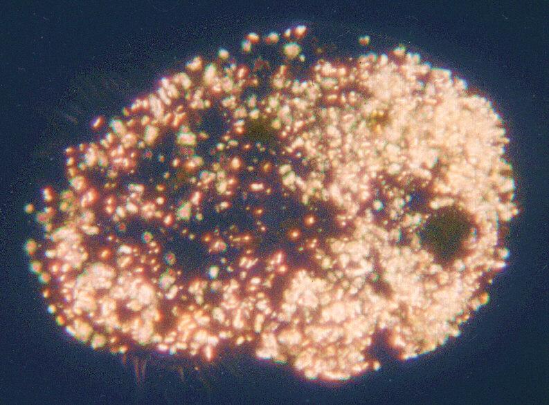
The second photo is rather an entertainment than an information shot. It
shows the same individual as the previous image, pictured through
crossed polarization filters. As I told you this is a way to visualize
crystallized substance which ciliates use to maintain concentration
equilibria of their protoplasma. I did a little colour saturation
enhancement to emphasize the typical effect of crystals being watched in
polarized light: they usually show up in rainbow colours as a result of
interference depending on the angle of the crystal axis to the axis of
the polarized light rays. In the phase contrast shot the crystals just
show up as dark-grey particles in Euplotes' protoplasma. This is typical
- there is hardly one single illumination method in light microscopy
which reveals all the secrets of the organism being watched. Phase
contrast comes very close to it; as you can see, the additional results
of polarized light or darkfield can sometimes be amazing.
Some more shots still to come.
Have a nice Sunday,
Ralf
Content-Type: image/jpeg; name="Euplotespha.jpg"
Content-Type: image/jpeg; name="Euplotespol.jpg" |
^o^
Animal Pictures Archive for smart phones
^o^
|
|