|
| Query: ciliate | Result: 2nd of 22 | |
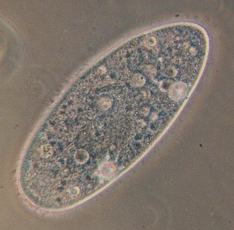
Protozoa - ciliates again - Paramecium caudatum
| Subject: | Protozoa - ciliates again - Paramecium caudatum
| | Poster: | Schmode (schmode@vossnet.de)
| |

| File size : 87002 bytes
File date : 2000:11:20 11:11:33
Resolution: 751x738
Jpeg process : Baseline
Posted Newsgroups: alt.binaries.pictures.animals
Posted Date: Sat, 16 May 1998 22:32:47 +0200 |
Hello everybody,
may I introduce: Paramecium caudatum.
This protozoon is known to me with its full name. If i just wrote
"ciliate" in my previous postings it was just to help out; protozoa,
especially ciliates, are sometimes hard to identify, and I just write
"ciliate" in these cases.
This one, however, has so many distinctive features that it is easy to
identify. Moreover, Paramecium caudatum (which even has a common german
name which would be about "houseshoe animal" in english) is the darling
of all protozoologists and microscopers. It hardly ever feeds on algae
but on bacteria which makes its body structure transparent; the bacteria
diet also makes it easily hatchable if you know how to supply tasty
bacteria (e-mail me in case you are interested in hatching Paramecium;
I'll tell you how).
Look at the big dark-grey oval in the lower middle of Paramecium; this
is the nucleus where the genetic information is stored. If you look
close you will see a darker-grey spot in the middle of the nucleus; this
is the nucleolus, a sub-nucleus which, as far as I know, comes into play
when sexual reproduction takes place. The banana-shaped, dark-grey zone
in the upper middle is the mouth; the bacteria are inserted here by
specialized cilia.
You can also see two big bright bubbles at the bottom side of
Paramecium; these are the excretion organs called "contractile
vacuoles". The waste liquids are delivered there by small channels
radially attached to the vacuole; you can guess them when you watch the
5 or 6 bright spots round the lower one of the two vacuoles. Please keep
that in mind if you watch my further postings because I have much better
shots of that. If you watch live Paramecia under the microscope you can
constantly see the vacuoles getting bigger, and about every minute one
of them contracts and expels its content outside Paramecium's body.
I should also mention the white, very small, sand-grain-like structures
you can see especially in the upper half of the body. I'll explain those
later; it's a bit complicated and it should be accompanied by a more
adequate shot which will be one of the next I post. Stay tuned, you'll
be surprised.
Have a nice sunday
Ralf
Content-Type: image/jpeg; name="Paramecium.jpg"
|
Comments |
|---|
| | your-name |
|
hi !
nice pict.
Do you know some good books about paramecia ?
best regards
pol |
| | hanie |
|
hi
i have a question do u khow some information about oligotrichia in cliates?
thank u |
| | maryam |
|
can you show me some pictures of various vacuole?
thank you |
| | "&;' |
|
| what is excertion of a paramecia |
^o^
Animal Pictures Archive for smart phones
^o^
|
|
|