|
| Query: Ciliate | Result: 13th of 22 | |
PING MacTrix - Protozoa - new scans, #5 - ciliate in excentric illumination
| Subject: | PING MacTrix - Protozoa - new scans, #5 - ciliate in excentric illumination
| | Poster: | Schmode (schmode@vossnet.de)
| |
| File size : 56727 bytes
File date : 2001:02:21 16:31:36
Resolution: 800x667
Jpeg process : Baseline
Posted Newsgroups: alt.binaries.pictures.animals
Posted Date: 17 Aug 1998 15:22:08 -0500 |
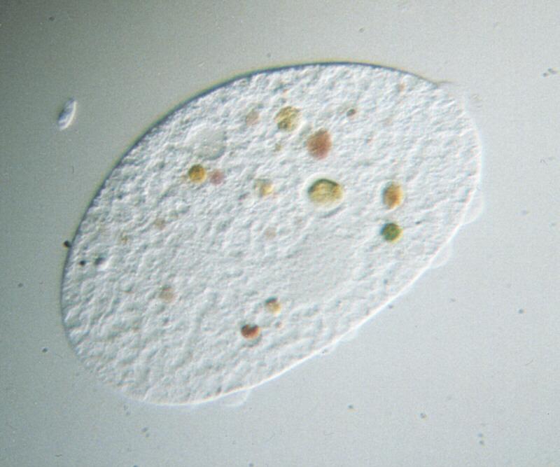
PING MacTrix - Protozoa - new scans, #5 - ciliate in excentric illumination
Hi again,
my yesterday?s posting featured an example of in what way illumination
can be used for enhancing the contrast of a microscopical image. I
showed you a Phacus oscillans in excentric illumination displaying its
flagellum which would not have been visible otherwise. I promised to
provide you with a shot of a ciliate made with the same method of
contrast enhancement.
That is what you are looking at right now. It shows a ciliate which I
have not yet been able to properly identify; it may be a member of the
Colpidium family. This organism would be almost totally transparent in
brightfield illumination in which the light source axis would be
parallel to the axis of the microscope lens and vertical to the object
slide.
Now see what excentric illumination does to that picture. The whole body
all of a sudden becomes structurized. The vacuole at the upper middle of
the body which is used for expulsion of waste liquids shows up like a
moon crater, the nucleus reveals its own grainy structure, and the outer
cell membrane looks a lot rougher than it is expected to be. Even the
algae being digested show a spherical apperance; being all of the same
species they nicely illustrate the colour change due to the progressive
process of degradation by the ciliate?s enzymes.
These contrasts, beautiful as they are, are the danger with excentric
illumination. Among the structures I mentioned the vacuole, the nucleus
and the algae are true, the roughness of the outer membrane is not -
have a look at the organism?s outline and you?ll realize. The roughness
actually comes from the structures inside the organism?s protoplasma.
The outer cell membrane itself has no changes in optical density
whatsoever, so it doesn?t show up with excentric as well as with
vertical illumination. The human
brain though knows there must be an outer coating of the organism, so
the contrasting structures deeper in the protoplasma are projected onto
and taken for the cell membrane. The phenomenon of ?false contrasts"
which can also occur with other illumination techniques is a major
problem in light microscopy; scientific microphotography hardly makes
use of excentric illumination for that reason.
I do and, hopefully, if time allows, will continue to make shots like
that. False or not, I like these contrasts.
See you,
Ralf
name="Ciliateexc.jpg" |
^o^
Animal Pictures Archive for smart phones
^o^
|
|
