|
| Query: protozoa | Result: 26th of 36 | |
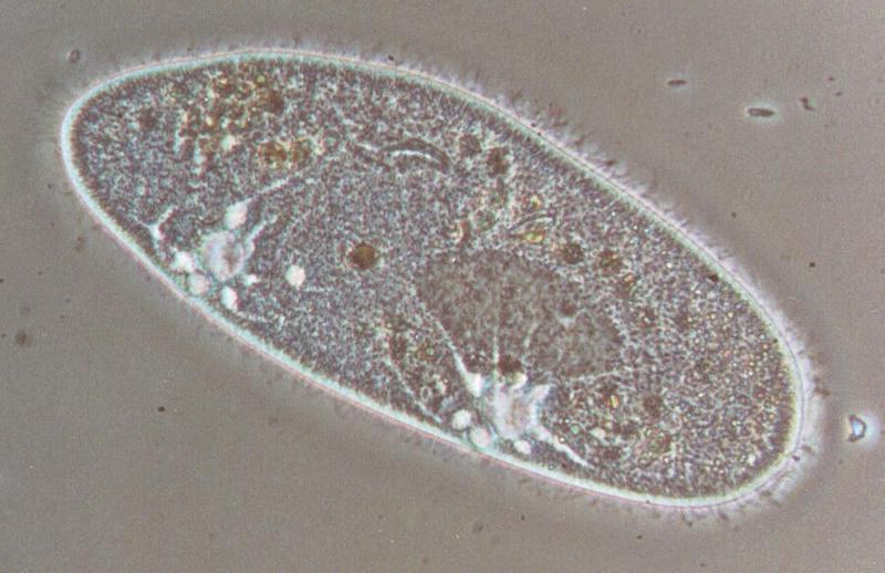
Protozoa - Paramecium caudatum take three - REPOST
| Subject: | Protozoa - Paramecium caudatum take three - REPOST
| | Poster: | Schmode (schmode@vossnet.de)
| |

| File size : 78979 bytes
File date : 2000:11:20 11:11:33
Resolution: 900x583
Jpeg process : Baseline
Posted Newsgroups: alt.binaries.pictures.animals
Posted Date: Mon, 25 May 1998 23:12:55 +0200 |
REPOST - This posting was usually scheduled for Saturday. Apparently the
news server didn't do what I wanted; I couldn't find my posting on any
other server till Monday. In order to make sure you can see this one
here comes a second try; sorry if you have already downloaded it.
Hi again,
Here comes my third shot featuring Paramecium caudatum.
Most facts I had to tell you about it have already been told in my
previous postings of that guy. Just have a look at the two contractile
vacuoles at the bottom of the picture. As I already told you these
organs manage the excretion of Paramecium; they are fed by radially
attached small channels which were hardly visible in my first Paramecium
posting but come out beautifully in this shot. Especially when you watch
the right of the two vacuoles you can see them like sunbeams pointing
away from the vacuole. The nucleus is also clearly visible in the right
half of the organism; the dark-grey spot attached to its top is the
nucleolus. Paramecium's mouth can be seen in the upper middle of its
body.
This photo nicely shows the location of Paramecium's cilia. You can see
that there is not only a ring of cilia round the organism's shape; if
you look closer it seems as if this one hadn't shaved for a couple of
days, meaning, of course, that Paramecium's whole body is covered with
cilia. This is not always the case; I'll show you a ciliate later with a
different kind and location of the cilia.
Hope you can bear the suspense.
Have a nice weekend!
Ralf
Content-Type: image/jpeg; name="Paramecium3.jpg" |
^o^
Animal Pictures Archive for smart phones
^o^
|
|

