|
| Query: Protozoa | Result: 20th of 36 | |
Protozoa series - new scans, #6 - a ciliate, darkfield and an off-topic bonus shot
| Subject: | Protozoa series - new scans, #6 - a ciliate, darkfield and an off-topic bonus shot
| | Poster: | Schmode (schmode@vossnet.de)
| |

| File size : 78079 bytes
File date : 2001:02:21 16:31:24
Resolution: 886x671
Jpeg process : Baseline
Posted Newsgroups: alt.binaries.pictures.animals
Posted Date: 16 Aug 1998 10:24:00 -0500 |
Protozoa series - new scans, #6 - a ciliate, darkfield and an off-topic bonus shot
Hello again,
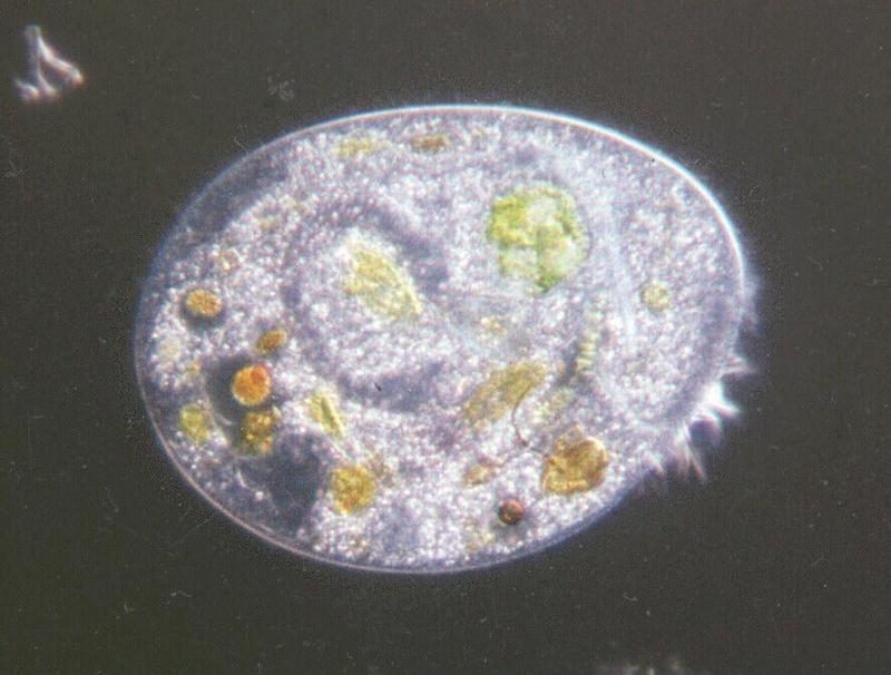
this shot is a little tricky. I like it a lot, but I do not know exactly
what it features. The organism pictured here definitely is a ciliate;
the location of the cilia suggests a climacostomum virens. If I am
right, the oval zone in the middle of the organism is not the nucleus;
it is the darker open ring around it - just compare to the Climacostum
virens posting I have made before. However, I may be wrong, as I may be
in some cases with my identifications. If there are any biologists out
there I would appreciate correction in case I have misidentified some of
the guys pictured here.
I promised to make you familiar with another technique off illumination
- here it comes. The medhod being in use here is called darkfield
illumination; it is somewhat the opposite of the method commonly used
which I already introduced as brightfield illumination which uses the
light passing through and contrast being based on absorption of the
object.
The principle of darkfield is quite simple: The light source of a
microscope is usually far away from the object in relation to the
object's size. That means the light rays reaching the object are almost
parallel. They are focused on the object by a lens system called
condensor which is between the light source and the object. If you
exclude all light rays which would directly reach the microscope lens
from being focused - an easy but effective way is to put a black plastic
disc in the center of the condensor - there is no direct illumination
and the scenery becomes completely dark. However, the light passing the
disc is focused on the object. The structures in the object vary in
refraction index, thus causing light flexion, total reflection and
interference, all these re-directing sufficient light into the
microscope lens to provide a beautifully contrasted image. Especially
the food vacuoles which manage algae digestion show up vividly coloured
and sharply contrasted. Darkfield illumination, by the way, makes best
use of the microscope's optical resolution because it uses the
peripheral light rays which in brightfield illumination have to be
excluded by closing the lens aperture to prevent over-illumination.
I have included another shot to show you what darkfield microscopy is
capable of revealing. SORRY, this is definitely off-topic - it is a
colony-forming alga called Volvox, almost 1/10 of an inch in diameter, a
giant sphere floating through the water actively propelled by flagella.
I have no more Protozoa shots using darkfield, so please forgive my
using a plantal image for further illustration of the method.
I haven't counted yet but some shots still to come.
Greetings from the Old World,
Ralf
name="Ciliatedark.jpg"
name="Volvox.jpg" |
^o^
Animal Pictures Archive for smart phones
^o^
|
|

