|
Protozoa series - new scans, #9 - a huge fat ciliate
| 제목: | Protozoa series - new scans, #9 - a huge fat ciliate
| | 올린이: | Ralf Schmode (schmode@vossnet.de)
| |

| 파일크기 : 85004 bytes
File date : 1998:08:26 09:00:00
해상도: 800x701
Jpeg process : Baseline
Posted Newsgroups: alt.binaries.pictures.animals
Posted 촬영일: 25 Aug 1998 18:01:28 -0500 |
Protozoa series - new scans, #9 - a huge fat ciliate
Hi everybody,
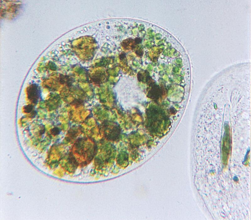
the shot I am bringing you today is a bit difficult. The organism being
pictured here is no doubt a ciliate; you can see a row of cilia around
the outline of its body.
The problem is that the water sample I found this guy in was an
extremely well fertilized ditch in an agricultural environment. I like
these when going on hunt for something to focus my microscope upon; the
conditions in these ditches with all the nitrogen in them are ideal for
algae as well as for bacteria - and for anyone who loves them for
dinner. Hungry ciliates are extremely numerous in such an environment,
so there is a good chance to get some nice shots when dealing with these
water samples.
The problem is that fat ciliates are almost impossible to identify. Look
at the one fully pictured here; it is so filled up with algae digestion
vacuoles that distinctive features are impossible to find. Apart from
the nucleus which you can guess as being the only colourless zone in the
ciliate's body there is nothing but different kinds of green. May be the
ciliate to its right which is incompletely pictured is a clue: It is on
a diet of just one diatomea - the greenish barrel in its protoplasma -
and it clearly displays its mouth funnel, allowing to identify it as a
Nassula. So, first guess for the fat guy is also Nassula; the mouth
funnel may be covered with digestion vacuoles. However, if you have a
close look at the outline of the organism you can see a tiny gap at the
front end; that would be typical to another ciliate called Prorodon
teres. If it is a Prorodon there should also be a mouth funnel at the
front end, behind the small gap; it may be invisible due to being
covered with algae being digested. By the way, the two ciliates pictured
here a of extraordinary size, each measuring about 1/50 of an inch in
length: ciliates, in general, even if belonging to the same species, are
very variable in size depending on their feeding condition.
I, personally, like this shot. Doesn't provide much detail but makes up
with lots of colour - one of the rare cases in which phase contrast
illumination would be inappropriate. Good old brightfield is just right
for this huge fellow.
The next picture I shall post will be phase contrast again. It has one
thing in common with today's feature ciliate: I do not know exactly what
it is.
So long
Ralf
Content-Type: image/jpeg; name="Fatciliate.jpg"
|
댓글 |
|---|
| | Nicelle Sernadilla, Editor |
|
Greetings!
I would like to request permission to use the image of a protozoa found in
the website
http://www.animalpicturesarchive.com/view.php?tid=1&did=50193
as follows:
Image placement: inside page
Image size: 1/16 A4
In the publications:
1. Biology: A Course for 'O' Level Textbook
(2nd edition)
2. Comprehensive Biology for 'O' Level
Science (2nd Edition)
These are price-controlled textbooks produced by Marshall Cavendish
International (Singapore) Private Limited, in collaboration with the
Ministry of Education, Singapore.
In view of the educational nature of the publications, we hope that you
will consider granting use of the image. We would of course fully credit
the photographer/website in the acknowledgements. Please let us know how
you would like to be credited.
Thank you. I look forward to hearing from you. |
^o^
동물그림창고 똑똑전화 누리집
^o^
|
|
|